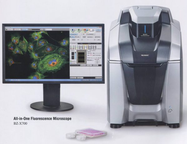
Keyence All-In-One Fluorescence Microscope
The fully enclosed stage chamber of this Keyence microscope allows all imaging to be done under normal room lighting conditions. The microscope is fully under computer mouse control for X,Y,Z stage, filter cube rotator and turret objective switching device. It automatically maps out viewing area for slides, dishes and titerplates. Filter cubes (4) are easily switched in and out during observations without the need to turn off the light. Standard configuration has DAPI, GFP, mRFP and an open position for brightfield but cubes are available for Cy5/iRFP (640 line on laser systems) and NeuO, a neuronal viability stain that is excited at 488 and emits at >620 nm. In brightfield mode, oblique illumination is used to achieve near DIC images. A stage incubator for temperature and humidity control is also available. Standard objectives (all Nikon) include 2x, 10x, 20x, 40x, 40x long working distance (LWD), and 100x oil. Although not a confocal system it performs image stacks at near confocal resolution with a minimum of 0.1 µm steps. It will obtain stacks through 500 µm thick slices. The unit has a high sensitivity cooled monochrome CCD that is converted to a 3CCD color camera for staining images at the click of a button. A large motorized stage, high-speed autofocus, low photobleaching mode and real-time image overlays are features of the core microscope. Advanced imaging functions that are all available include image stitching in either an autofocus mode for each image (slow) or full focus wide-area (about 30 sec to scan a single plane human hippocampal slice in brightfield and display the montage), navigation (maintains image of montage on side panel and lets you see on the montage where you are located for higher magnification imaging), optical sectioning, and time-lapse. A structured illumination module use projection of an optical grid to achieve super-resolution fluorescence images.
A Peltier cooled stage adapter is available for use on this microscope. It will allow imaging of cells in 35 mm glass bottom dishes (without the double thick plastic walls at the base). This device will cool cells in medium at approximately -20oC/min to below 0oC, although the precise rates of cooling need to be determined for the volume of medium. Continuous fluorescence imaging (time lapse/multi point) can take place while cooling. Training is required for this application since no specific stage adapter is yet available.
Analysis software is included on the system and an off-line package is also available on a second computer in the facility. Applications include hybrid cell counts (can use dual fluorescence or brightfield/fluorescence to automatically quantify the % of cells expressing tags along with color extraction (for stained sections) and masking functions for selection of regions of interest. A macro cell count application allows parameters set for one acquisition to be applied to multiple samples, especially useful for batch processing and high throughput analysis. A measurement module allows point and click 2D measurements for length, distance, area, etc. and a 3D module allows visualization fluorescence image stacks with rotation, zoom in and out functions, sectional views, XYZ slicing and maximum projections.
Microscope Information
Location
Room 224 MRB
Costs
$4/ hr or $30/ month if used over 7.5 hrs in a calendar month.
Supervisor and Training
Alisa Shaw:
[email protected]
Phone: 491-5531
Until July 1, 2019, preference for use will go to major users of the grant application that secured the microscope funding. They will be allowed reservations up to 10 days in advance. Booking for others will open 3 days in advance.
Online Booking is for trained individuals only, and can be accessed here.